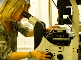
The blood vessel tissue is examined using phase-contrast microscopy, which makes it possible to follow the movements in the tissue, before, during and after cell division.
University Copenhagen’s Niels Bohr Institute research demonstrates that cell division is very ordered
Published 9, December 2014 at 275 × 207 in University Copenhagen’s Niels Bohr Institute research demonstrates that cell division is very ordered
Bookmark the permalink.

