
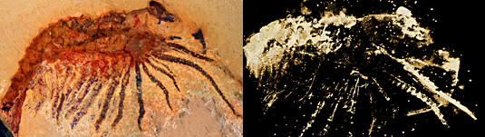
The figure on the left shows a light micrograph of the fossil, while the microtomographic image right reveals fine details of structures hitherto concealed within the slab.
The figure on the left shows a light micrograph of the fossil, while the microtomographic image right reveals fine details of structures hitherto concealed within the slab.