
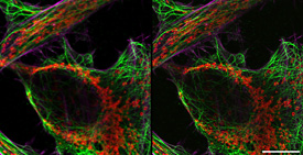
Comparison between confocal and Airyscan image. HeLa cells, red: mitochondria membrane, green: microtubuli, magenta: actin fibres; acquired with LSM 800. Scale bar 5 µm. Sample courtesy of A. Seitz, EPFL, Lausanne, Switzerland.
Comparison between confocal and Airyscan image. HeLa cells, red: mitochondria membrane, green: microtubuli, magenta: actin fibres; acquired with LSM 800. Scale bar 5 µm. Sample courtesy of A. Seitz, EPFL, Lausanne, Switzerland.